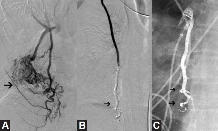
Figure 3.

(A-C): Selective right internal mammary artery injection (A) shows the presence of a high-flow right internal mammary artery (IMA) to pulmonary artery malformation (arrow). Selective right IMA arteriogram (B) following coil occlusion of the IMA distal to the lowest contributory branch and Onyx injection shows cessation of flow to the malformation (arrow). Radiograph of the chest (C) the day after embolization shows penetration of Onyx (arrows) into the first-order branches of the IMA
