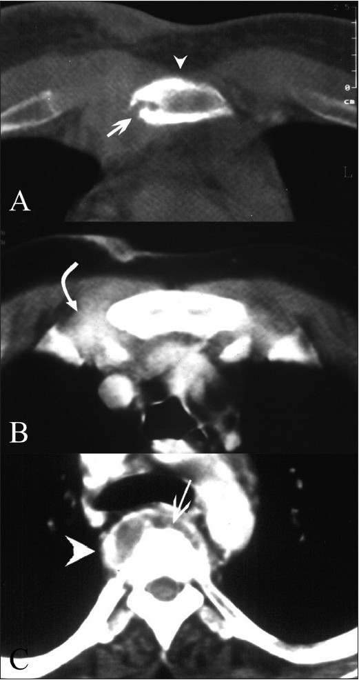
Figure 2 (A–C).

Sternal tuberculosis – patient 1. Contrast-enhanced CT scans show erosion (arrow) and sclerosis (arrowhead) of the sternum. The adjoining right parasternal soft tissue shows thickening with dense enhancement (curved arrow). Vertebral destruction (arrow in C) and a paravertebral abscess (arrowhead in C) are also noted
