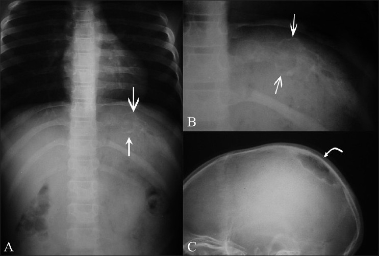
Figure 4 (A–C).

Rib tuberculosis – patient 8. Frontal radiographs of the ribs (A, B) show an expansile osteolytic lesion with sclerosis involving the posterior end of the left 10th rib (arrows). Lateral skull radiograph (C) of the same patient shows a sharply marginated osteolytic lesion, with marginal sclerosis in the right parietal bone (curved arrow)
