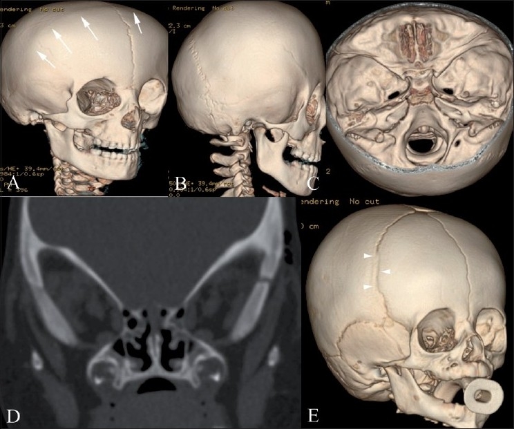
Figure 3(A-E).

Bilateral (A-D) and unilateral partial (E) coronal synostosis 3DCT volume rendered images (A-C, E) and coronal CT scan (D). There is complete fusion of the coronal suture (white arrows) with a prominent frontal bone and flattened occiput. Coronal reconstruction (D) demonstrates prominent bilateral elliptical orbits, known as the “harlequin eye” deformity. Note the early partial fusion of the right coronal suture (arrowheads in E)
