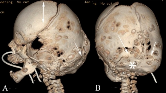
Figure 5(A,B).

Turricephaly. Lateral (A) and posterior (B) 3DCT volume rendered images show turricephaly secondary to bilateral lambdoid fusion (arrows). Note the small, underdeveloped posterior fossa (*), and the “tall” cranium (double-headed arrow)
