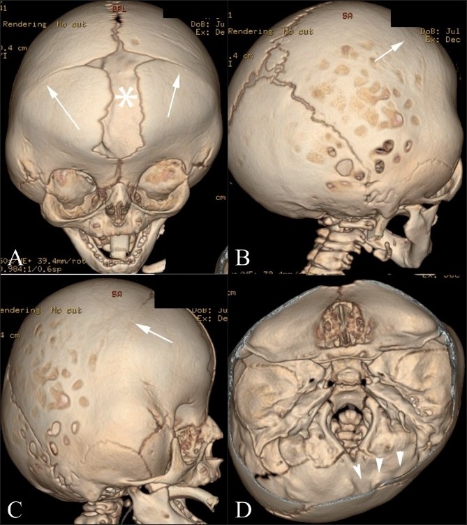
Figure 13(A-D).

Apert’s syndrome. Frontal (A), lateral (B), right posterolateral (C) and endocranial (D) 3DCT volume rendered images show coronal synostosis (arrows), gaping frontal midline defect (FNx01), and a small malformed skull base (arrowheads)
