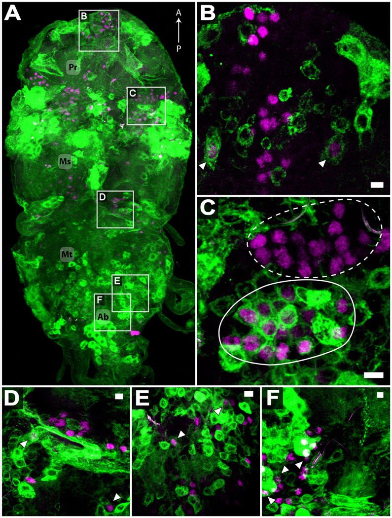
Figure 5.
Immunocytochemistry of adult w; tshGAL4, UAS-mCD8-GFP/CyO CNS, visualizing endogenous, membrane bound GFP (green) and FruM immunoreactivity (magenta). (A) Dorsal-ventral view of adult ventral ganglia. Extensive labeling is visible in prothoracic (Pr), mesothoracic (Ms), metathoracic (Mt), and abdominal (Ab) segments. Anterior-posterior axis is indicated. (B–F) 3 – 5 µm representative sections of the five groups of fruM neurons in the ventral ganglia, according to (Lee et al., 2000). FruM neural cluster 16 (B), 17 (C), 18 (D), 19 (E), and 20 (F). Arrowheads indicate examples of neurons coexpressing FruM and mCD8-GFP. In (C), a FruM-expressing cluster clearly coexpresses GFP (solid line), while an adjacent FruM cluster does not (dashed line). Scale bars (B–F) represent 5 µm.

