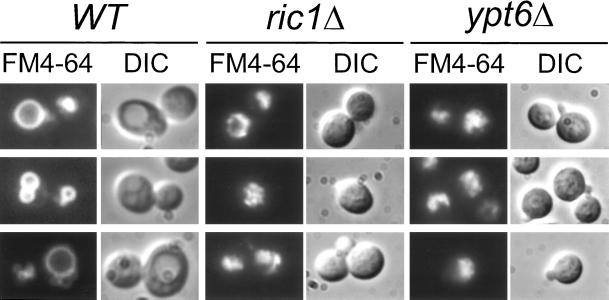
Figure 5.
ric1Δ and ypt6Δ cells display fragmented vacuoles. Wild-type (WT, SEY6210), ric1Δ (GPY1480), and ypt6Δ (GPY1700) strains were grown overnight in SDYE media, at 30°C, to midlogarithmic phase. Cells were incubated with FM4–64 for 15 min, resuspended in fresh media devoid of dye, and allowed to internalize the dye for 45 min, at 30°C, before viewing. FM4–64, fluorescence (left); DIC, differential interference contrast optics (right).

