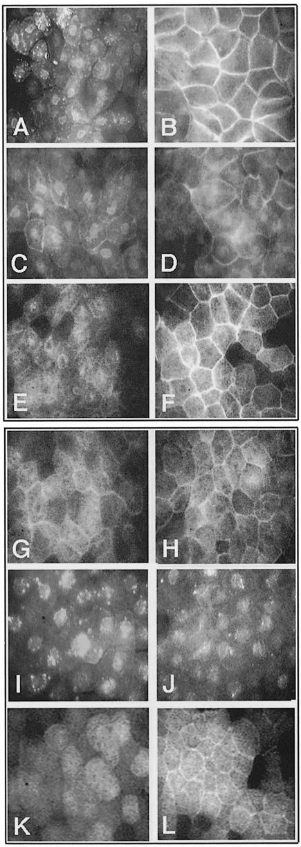
Fig. 4. Translocation abilities of Xdsh–GFP deletions upon rat frizzled-1 co-expression: the C–terminal DEP domain-containing region is necessary and sufficient for Xdsh membrane translocation. (A) Xdsh localizes in various subcellular regions of animal cap cells in punctuate appearance resembling vesicular structures. These are distributed in the cytoplasm, at the membrane and around the nucleus. (B) Upon frizzled co-expression, Xdsh is localized prominently at the membrane. Almost no Xdsh is seen in cytosolic locations. (C) D1, which lacks much of the DIX domain and flanking regions, is distributed similarly within the cell and shows less obvious accumulation than Xdsh in vesicles. (D) D1 translocates poorly to the membrane upon frizzled co-injection. (E) D2, which lacks the entire PDZ domain, is distributed in the entire cell, and (F) upon frizzled co-injection is localized prominently to the membrane. (G) D4, which lacks half of the PDZ domain, is distributed evenly in cells; (H) frizzled co-expression can mediate its membrane localization only to a very small extent. (I) D6 lacks the C–terminal region containing most of the DEP domain and localizes prominently to punctuate accumulations within the cells. (J) This cellular distribution of D6 does not change upon frizzled injection, and no membrane localization is observed. (K) D9, containing only the C–terminal region of Xdsh encompassing the DEP domain, shows mainly a cytoplasmic distribution. (L) frizzled co-expression clearly can promote D9 accumulation at the membrane.

An official website of the United States government
Here's how you know
Official websites use .gov
A
.gov website belongs to an official
government organization in the United States.
Secure .gov websites use HTTPS
A lock (
) or https:// means you've safely
connected to the .gov website. Share sensitive
information only on official, secure websites.
