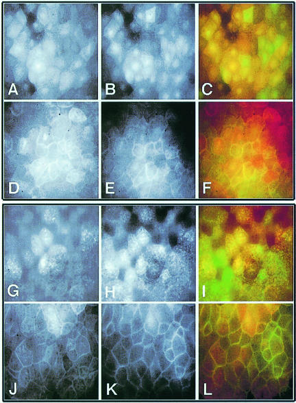
Fig. 7. Homomeric complex formation of Xdsh: membrane co-translocation of translocation-defective deletions with wild-type Xdsh. mRNAs for Xdsh-HA (A and D) and D6–GFP (B and E) were co-injected in early stage embryos without (A–C) or with rat frizzled-1 mRNA (D–F). The subcellular location of mutant and wild-type Xdsh in gastrula animal caps is shown. Images on the left (A and D) show the fluorescence of rhodamine-labeled Xdsh-HA (red channel), middle images (B and E) show the GFP fluorescence of D6–GFP (green channel) in the same animal cap tissue; and images on the right (C and F) are overlays of both signals. Note that D6–GFP (E) is found associated with the membrane when it is co-injected with Xdsh-HA and frizzled, while it remains cytoplasmic/vesicular when co-injected with frizzled alone (compare Figure 4J). D8-HA (G and J) co-injection with Xdsh–GFP (H and K) results in D8-HA accumulation at the membrane, when co-injected with frizzled (J–L). D8 on its own with frizzled does not localize well to the membrane (data not shown). Left panels (red channel) are Texas red signals of D8-HA (G and J); middle panels (green channel) are GFP signals (H and K) and right panels are overlays of both signals (I and L).

An official website of the United States government
Here's how you know
Official websites use .gov
A
.gov website belongs to an official
government organization in the United States.
Secure .gov websites use HTTPS
A lock (
) or https:// means you've safely
connected to the .gov website. Share sensitive
information only on official, secure websites.
