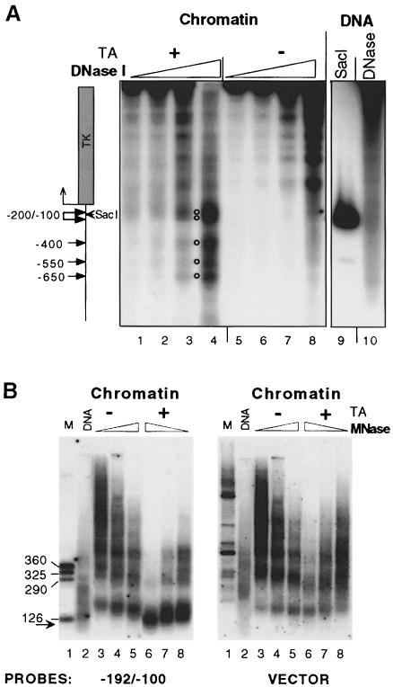
Fig. 2. Chromatin structure of the MMTV promoter. (A) Hormone-dependent DNase I-hypersensitive sites are located in the MMTV LTR. Groups of 12 oocytes were injected with 1 ng of pMTV:M13 coding ssDNA, 5 ng of dsDNA for pSTC GR 3-795 and 0.25 ng for pAdML reference (lanes 1–8). After overnight incubation, hormone was added (TA; 1 μM) (lanes 1–4) or not added (lanes 5–8) and oocytes were harvested after 24 h for the DNase I hypersensitivity assay. Lane 9, internal molecular weight marker showing the position of the SacI restriction enzyme cut. Lane 10, naked dsMMTV promoter DNA digested with DNase I. (B) MNase in situ digestion shows hormone-dependent disruption of the canonical nucleosome structure in the vicinity of GRE elements. Groups of 10 oocytes were injected. The next day, hormone (TA; 1 μM) was added as indicated and oocytes were harvested after 24 h for MNase digestion. DNA was resolved in an agarose gel, transferred and hybridized with a labeled MMTV promoter probe encompassing region –192/–100, and then washed and rehybridized with an M13 vector probe. Lane 1, internal DNA marker; lane 2, naked dsMMTV promoter DNA digested with MNase. The arrow shows a subnucleosomal particle ∼120 bp DNA fragment revealed only after hybridization with specific probe.

An official website of the United States government
Here's how you know
Official websites use .gov
A
.gov website belongs to an official
government organization in the United States.
Secure .gov websites use HTTPS
A lock (
) or https:// means you've safely
connected to the .gov website. Share sensitive
information only on official, secure websites.
