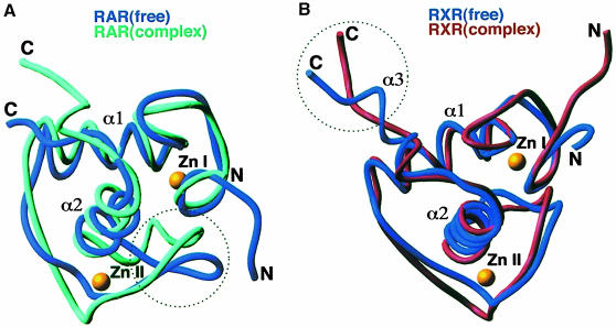
Fig. 6. Protein structural transitions induced in the RXR–RAR–DR1 complex. (A) The superposition of the free RAR DBD structure derived from NMR studies with its structure in the RXR–RAR–DR1 complex. The dotted circle indicates that a major structural rearrangement induced on assembly occurs along the Zn–II loop. (B) The superposition of the free RXR DBD structure from NMR studies with its structure in the RXR–RAR–DR1 complex. The dotted circle indicates that a major structural reorganization occurs along the α3 helix of the T–box, which is disrupted in forming the complex.

An official website of the United States government
Here's how you know
Official websites use .gov
A
.gov website belongs to an official
government organization in the United States.
Secure .gov websites use HTTPS
A lock (
) or https:// means you've safely
connected to the .gov website. Share sensitive
information only on official, secure websites.
