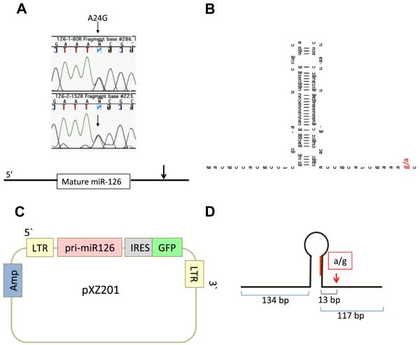
Fig. 1.
A base substitution in pri-miR-126 in RS4;11 cells is identified. (A) DNA chromatograms illustrating the genomic sequence of miR-126 DNA from RS4;11 cells. Nucleotides are indicated by capital letters. The green, black, and blue wave indicates A, G, and C, respectively. The arrows indicate the base position of A24G base substitution. Note that G and A sequence are both present. The base substitution is located 24 bp from the 3′end of mature miR-126 miRNA. (B) The location of the base substitution in the pri-miR-126. The red capital indicates A24G. (C) The structure of the pXZ201-pri-miR-126. Pri-miR-126 genomic sequence was cloned into the pXZ201 retroviral expression vector. (D) The structure of the pri-miR-126 cloned from the genome of RS4;11 cells. 5′ and 3′ flanking regions was 134 and 117 base long, respectively, which are enough length for processed from pri-miRNA to pre-miRNA [27].

