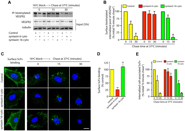
Figure 4.
Syntaxin 6 inhibition does not target endocytic pool of VEGFR2 for degradation. (A-E) Serum-starved uninfected (Control) and syntaxin 6-cyto or syntaxin 16-cyto infected HUVECs (20 hours of infection). (A-B) Cells were surface biotinylated as described for Figure 1F. Samples were then incubated at 16°C for 30 minutes to allow biotinylated VEGFR2 to accumulate in endosomes. The remaining surface biotin was removed by treatment with sodium 2-mercaptoethanesulfonate (MesNa) and quenching of MesNa with iodoacetic acid. These samples (0-minute chase time point) were further incubated at 37°C for the indicated periods. To determine the levels of intracellular biotinylated VEGFR2 after different chase periods, cells were again treated with MesNa to remove any additional biotinylated VEGFR2 that had recycled to the cell surface. Intracellular biotinylated VEGFR2 was pulled down by streptavidin-Sepharose as described for Figure 1F. Levels of intracellular biotinylated VEGFR2 were quantitated by immunoblotting for VEGFR2. Total cell lysates were also immunoblotted with the indicated antibodies to assess relative protein abundance. (B) Densitometric analysis of immunoblots in panel A. Values are expressed relative to 0-minute chase time point (normalized to 100%). Values are presented as mean (± SD) for n = 3, P ≤ .05. (C,D) HUVECs were labeled with an E-tagged anti-VEGFR2 antibody (ScFv), at 10°C for 30 minutes, and then incubated with VEGF for an additional 30-minutes. Cells were washed and imaged to assess ScFv-binding at the cell surface. Samples in which ScFv was bound to the cell surface were further incubated at 16°C for 30 minutes. The remaining surface-bound antibody was removed by washing with low pH buffer. These samples (0-minute chase time point) were subjected to chase at 37°C in the presence of VEGF. Cell surface–localized and internalized ScFv-VEGFR2 complexes were detected using an FITC-conjugated antibody against the E-tag. (D,E) Graphs represent quantitative measurements of cell surface–localized and total internalized VEGFR2, as determined by image analysis. Values in panel E are expressed as the percentage of total internalized ScFv fluorescence at the 0-minute chase time point. Values in panels D and E represent mean (± SD) for n = 90 cells for each condition from 3 separate experiments, P ≤ .005. Scale bar represents 5μm.

