
Figure 13.6.
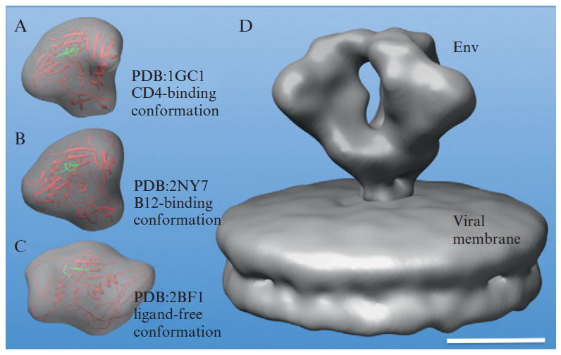
3D density map of HIV Env at 2.0 nm resolution (Liu et al., 2008). Comparison of EM map (D) with simulated maps calculated from three gp120 monomer crystal structures: CD4-binding conformation (A), b12-binding conformation (B), and ligand free conformation (C) indicates that the molecular docking remains challenging at 2.0 nm resolution. The scale bar is 5 nm.