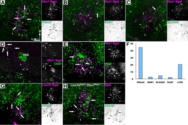
Figure 2.
Ventral medullary respiratory glutamatergic neurons are Dbx1 derived. A–E, G, H, Pseudocolor mosaic images of β-Gal (magenta) IHC from Dbx1LacZ/+ (A–E), Lmx1bLacZ/+ (G), or Dbx1LacZ/+;Lmx1bLacZ/+ double heterozygous (H) mutant mice with bright-field ISH (green) for VGlut2 (A, G, H), GAD1 (B), Slc6a5 (GlyT2) or (C), or IHC (green) for ChAT (D) or S100 (E) within the preBötC (A–C, E) or adjacent to the cNA (D). Lateral images correspond to single-channel images. F, Percentage of β-Gal cells in the VLM that coexpress specific genes. Arrows point out coexpression. Arrowheads indicate absence of coexpression. Scale bar, 200 μm. D, Dorsal; L, lateral; cNA, compact formation of nucleus ambiguus.

