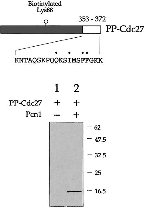
Fig. 4. In vitro Pcn1 binding assay using biotinylated PP–Cdc27 fusion protein (PP–Cdc27). Upper part: schematic representation of PP–Cdc27 showing the location of the biotinylated lysine residue and, at the C–terminus, residues corresponding to 353–372 in Cdc27. Lower part: following mixing of the PP–Cdc27 protein with recombinant Pcn1, proteins binding to Pcn1 were isolated by Ni–NTA affinity chromatography and subjected to SDS–PAGE. The bound proteins were then transferred to a PVDF membrane and probed using streptavidin-labelled alkaline phosphatase to detect the presence of the biotinylated PP–Cdc27 protein. Retention of the PP–Cdc27 protein was dependent upon the presence of Pcn1 (lane 2). See the text for details.

An official website of the United States government
Here's how you know
Official websites use .gov
A
.gov website belongs to an official
government organization in the United States.
Secure .gov websites use HTTPS
A lock (
) or https:// means you've safely
connected to the .gov website. Share sensitive
information only on official, secure websites.
