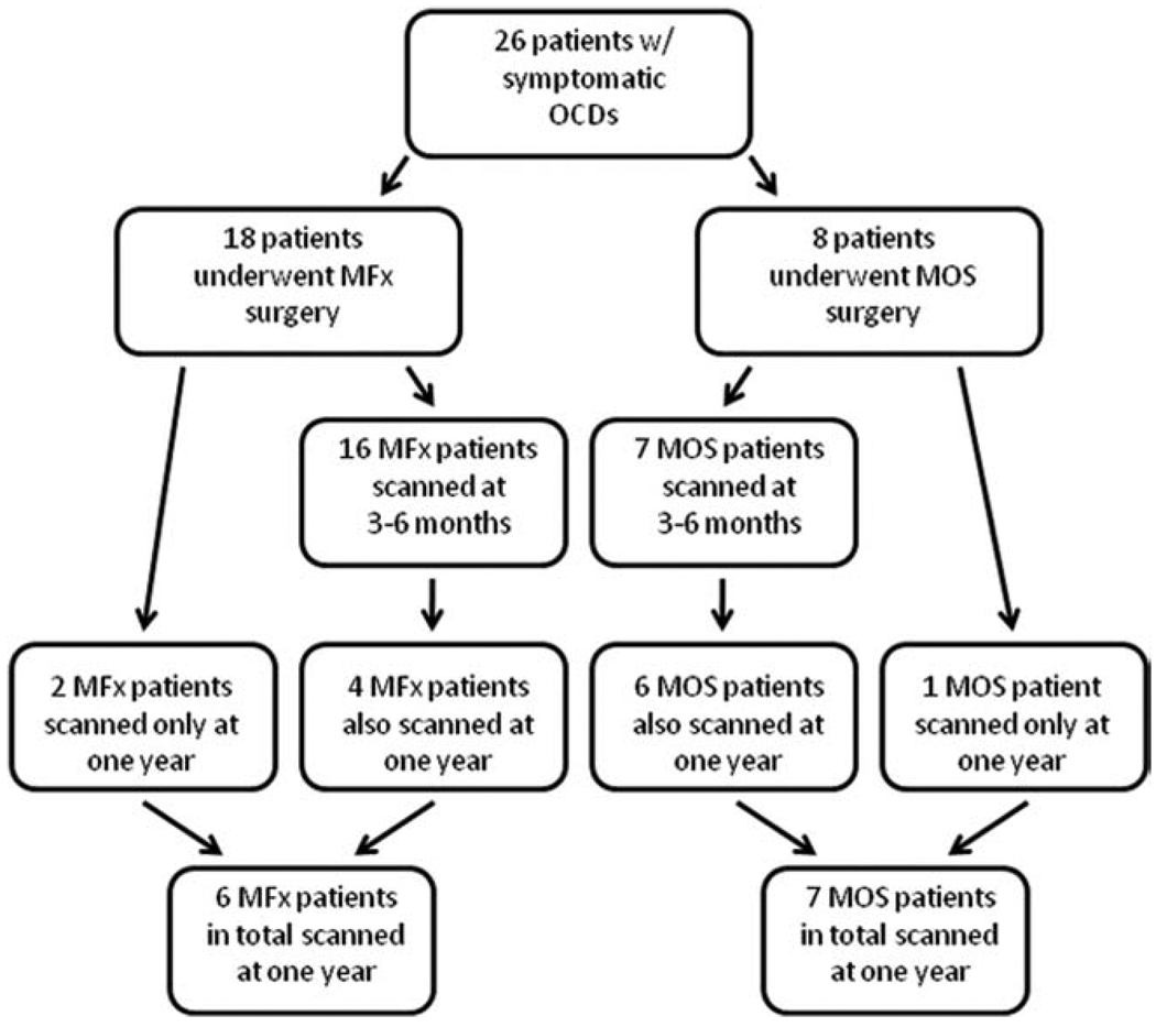
Figure 1.
Flow diagram detailing patients scanned for this study. Sixteen patients who underwent MFx were scanned at 3–6 months. In total, six MFx patients were scanned at 1 year. Seven patients who underwent MOS were scanned at 3–6 months. In total, seven MOS patients were scanned at 1 year. Data is nonrandomized, was gathered prospectively, and was analyzed cross-sectionally. CDs, focal cartilage defects; MFx, microfracture; MOS, mosaicplasty. [Color figure can be viewed in the online issue, which is available at wileyonlinelibrary.com.]

