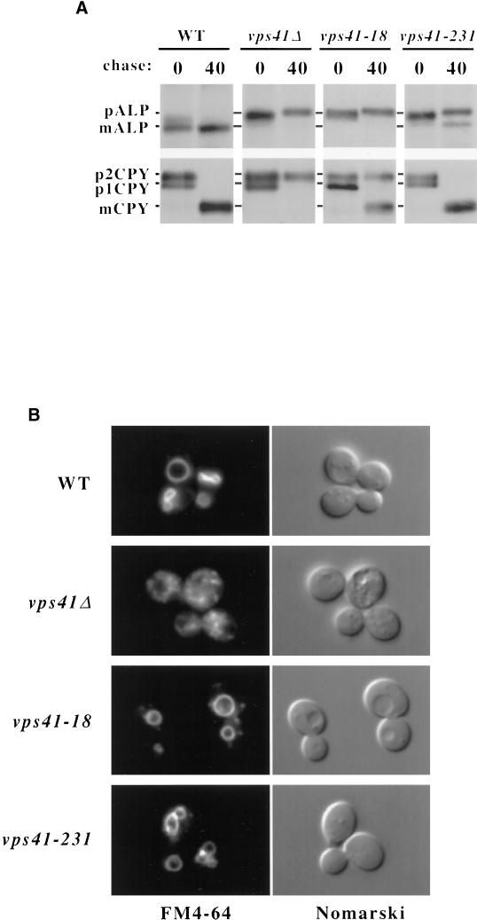
Figure 2.
Vacuolar protein sorting and vacuolar morphology of VPS41 mutants. (A) vps41Δ (WSY41) cells transformed with either complementing VPS41 plasmid (pVPS41), or plasmids containing vps41-18 (pVPS41-18), or vps41-231 (pTD44), were pulse-labeled with [35S]cysteine/methionine and chased for 40 min. CPY and ALP were immunoprecipitated with polyclonal antibodies and analyzed by SDS-PAGE and autoradiography (B). The same strains shown in A were labeled with FM4-64 for 15 min at 30°C and then chased in fresh media for 1 h at 30°C.

