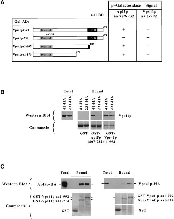
Figure 5.
Analysis of Vps41p interactions. (A) GAL4-BD fusion with a C-terminal fragment of Apl5p (aa 729–932) and a GAL4-BD fusion with full-length Vps41p tested against the indicated Vps41p fragments fused in frame with the GAL4-AD. β-Galactosidase assays were done in duplicate on multiple transformants for each experiment. (B and C) The indicated GST fusion proteins were purified from E. coli on GSH-Sepharose. (A) Triton X-100–solubilized 13,000 × g supernatant fractions from yeast cells expressing either Apl5p-HA (PRY1), Vps41p-HA (DKY25), or Vps41-231-HA protein (TDY30) were incubated with the immobilized GST fusion proteins at 4°C and then washed. The bound proteins were eluted with sample buffer. For Western blotting, 20% of the eluate was loaded per lane. One percent of solubilized extract (total) was loaded as a reference. (B and C) Top panels: a Western blot probed with anti-HA antibodies; bottom panels: Coomassie-stained gels of 10% of the eluted sample indicate the relative amounts of the fusion proteins used in each experiment. The identity of each GST-fusion protein is indicated to the right of the bottom panels.

