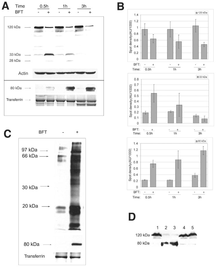
Fig. 1.
BFT induces release of the E-cadherin ectodomain and shedding of IEC membrane proteins. (A) BFT induces release of the E-cadherin ectodomain. Upper panel. Cell lysates of HT29/C1 cells treated with BFT (5 nM) for 0.5 hour to 3 hours were evaluated by western blot using the E2 antibody that recognizes the C-terminal domain of E-cadherin. Actin served as an internal control for protein loading. Blot is representative of five experiments. Lower panel. TCA-precipitated cell culture supernatants of HT29/C1 cells treated with BFT (5 nM) for 0.5 hour to 3 hours were evaluated by western blot using the H108 antibody that recognizes the ectodomain of E-cadherin. Transferrin, detected by Coomasie Blue staining, served as an internal control for protein loading. Blot is representative of three experiments. (B) Normalized western blot data demonstrating significant BFT-induced cleavage of intact E-cadherin (120 kDa) on HT29/C1 cells by 0.5 hour (P<0.01) with enhanced detection of a 33 kDa cell-associated E-cadherin fragment at 0.5 hour and release of the 80 kDa E-cadherin ectodomain into cell supernatants (both P<0.001). The 33 kDa E-cadherin fragment is degraded over time (see also Fig. 1A). Data are means ± s.d. of three experiments. (C) BFT (5 nM, 1 hour) induces shedding of IEC membrane proteins as well as the E-cadherin ectodomain (80 kDa). HT29/C1 cell membrane proteins were biotin-labeled and processed as in the Materials and Methods. Transferrin stained by Coomasie Blue serves as an internal control for protein loading. Blots are representative of four experiments. (D) Cleavage of E-cadherin extracellular domain requires biologically active BFT. HT29/C1 cells were treated with purified BFT (5 nM) or culture supernatants of B. fragilis 9343(pFD340::P-bft) that expresses wild-type BFT or B. fragilis 9343(pFD340::P-bftΔH352Y) that expresses mutant biologically inactive BFT. Cell lysates and TCA-precipitated cell culture supernatants were assessed by western blot using the E2 (upper lane) and H108 (lower lane) antibodies to the E-cadherin C-terminus or ectodomain, respectively. Lane 1, untreated control; lane 2 purified BFT; lane 3, B. fragilis 9343(pFD340::P-bft); lane 4, B. fragilis 9343(pFD340::P-bftΔH352Y); lane 5, brain heart infusion broth alone.

