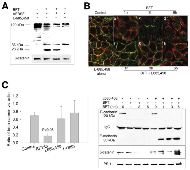
Fig. 3.
Inhibition of γ-secretase, but not β-secretase, blocks BFT-induced cleavage of the C-terminal domain of E-cadherin and partially β-catenin redistribution. (A) Cell lysates of HT29/C1 cells were analyzed by western blot using the E2 antibody to the C-terminus of E-cadherin. Cells were treated with BFT alone (30 minutes, 5 nM) or with inhibitors [β-secretase inhibitor, AEBSF (0.5 mM) or γ-secretase inhibitor, L-685,458 (1.5 μM)] for 30 minutes prior to BFT treatment. Molecular size markers indicate the 120 kDa intact E-cadherin and the 33 and 28 kDa degradation fragments of E-cadherin. β-catenin serves as an internal control for protein loading. Blot is representative of three experiments. (B) HT29/C1 cells were treated with BFT alone (5 nM) or in the presence of the γ-secretase inhibitor, L-685,458 (1.5 μM), followed by immunostaining with an anti-β-catenin antibody (CAT-5H10) (green) and the E2 antibody to the C-terminus of E-cadherin (red). Untreated control cells or cells pretreated for 30 minutes with L-685,458 (Panels a and e, respectively) reveal co-association (yellow) of E-cadherin and β-catenin staining. After BFT treatment, there is diffusion and dissociation of E-cadherin and β-catenin staining at 1 hour (Panel b) with progressive cytoplasmic β-catenin and E-cadherin diffusion over the subsequent 3- and 6-hour time points (Panels c,d). In cells pretreated with L-685,458, there is also dissociation of E-cadherin and β-catenin staining at 1 hour but the β-catenin signal is intense and localized at the cell membrane (Panel f). Over time, partial cytoplasmic diffusion of β-catenin occurs but with retention of a distinct β-catenin pool on the cell membrane (panels g,h). Representative of three experiments. (C) β-catenin/actin ratios in control HT29/C1 cells or cells treated with BFT (5 nM) for 6 hours in the presence or absence of L-685,458 (1.5 μM). L+B6h=L-685,458 + BFT for 6 hours. P<0.05, Control vs BFT, 6 hours. Data are means ± s.d. of five experiments (D) HT29/C1 cells were treated with BFT (5 nM) for 1, 3 or 6 hours in the presence or absence of L-685,458 (1.5 μM). Cell lysates were immunoprecipitated using an antibody to γ-secretase and the western blot analyzed using antibodies to the C-terminus of E-cadherin (E2) (upper panel), β-catenin (CAT-5H10) (middle panel) or γ-secretase (lower panel). Intact E-cadherin is 120 kDa. γ-secretase inhibition results in stable association of a 33 kDa C-terminal E-cadherin fragment and β-catenin with γ-secretase. Western blot is representative of two experiments.

