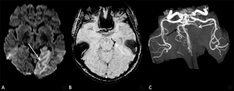
Figure 3 (A–C).

A 56-year-old female with acute stroke in the left PCA territory. Diffusion-weighted image (A) shows an acute infarct (arrow) in the left PCA territory. SWI image (B) shows blooming (arrow) in the P2 segment of the left PCA. TOF MRA (C) confirms thrombus (arrow) in the left PCA.
