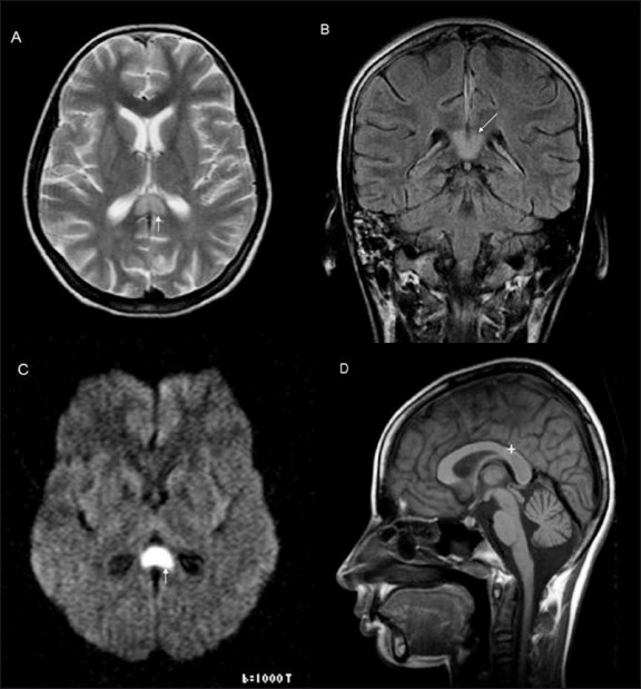
Figure 1 (A–D).

Axial T2W (A), coronal FLAIR (B), and diffusionweighted (C) images show a hyperintense, well-defined lesion (white arrow) in the splenium of the corpus callosum with a thin rim of surrounding non-hyperintense white matter. The same lesion on the sagittal T1W image (D) shows hypo to isointense signal (star)
