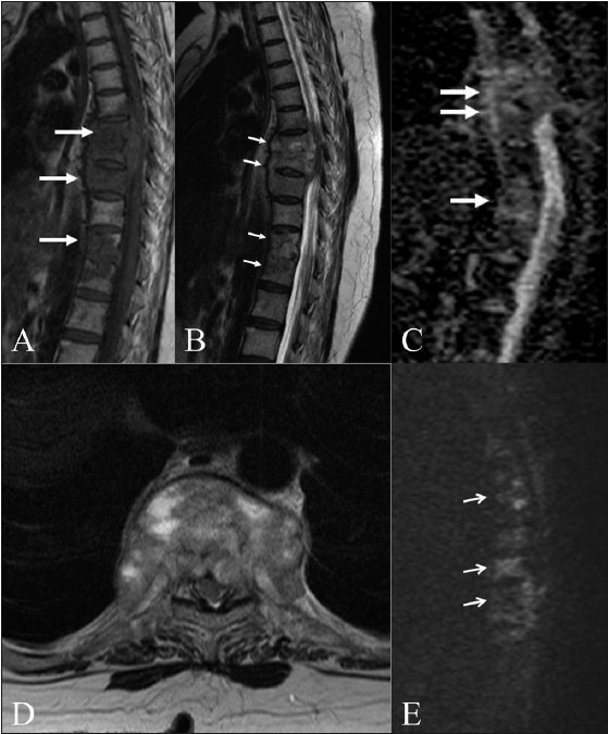
Figure 2 (A–E).

Tuberculosis of the spine. Sagittal T1W (A) and T2W (B) images in an elderly patient with tuberculosis show multifocal dorsal vertebral body involvement (arrows) with an epidural soft tissue component. Sagittal ADC map (C) and axial T2W (D) and diffusion (E) images show increased diffusion (arrows) in the involved vertebrae. (ADC: 1.42–1.5 × 10⁻³ mm2/s)
