Figure 6.

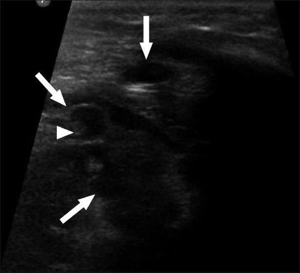
USG of the heart shows multiple cystic anechoic lesions (arrow) in the myocardium with a tiny hyperechoic scolex (arrowhead) within one of the cysts

USG of the heart shows multiple cystic anechoic lesions (arrow) in the myocardium with a tiny hyperechoic scolex (arrowhead) within one of the cysts