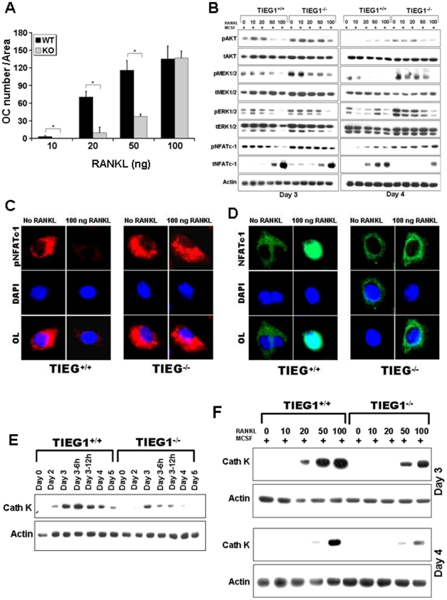
Figure 3. Differentiation defects in TIEG1−/− osteoclast lineage cells results from defective RANKL responses.
A. Mean number of osteoclasts from WT and TIEG1−/− (KO) mouse marrow following 4 days of culture in the presence of 25 ng/ml MCSF and the indicated concentrations of RANKL as outlined in the Methods section. Cells were stained and the number of osteoclasts quantitated as in Figure 1 (*p<0.05). These data were obtained from three replicate wells (p<0.05) and are representative of three separate experiments. B Osteoclast precursors at day 3 and mature osteoclasts at day 4 were treated with the indicated concentration of RANKL and equal amounts of total protein were analyzed by western blotting for the indicated phospho- or total proteins. These data are representative of two separate experiments. For each experiment, marrow cells from three mice were pooled and analyzed in three replicate wells. C and D. Confocal images demonstrating the effect of RANKL on phospho- NFATc1 (C) or total NFATc1 (D). Precursors were cultured with MCSF with or without RANKL as indicated. On day 3, cells were fixed and stained with the indicated primary antibodies. These data are representative of two separate experiments and each experiment is analyzed in three replicate wells. E and F. Time course expression of cathepsin K in WT and TIEG1−/− (KO) osteoclast precursors (day 3) and mature osteoclasts (day 4) cultured in the presence of MSCF alone (E) or with the indicated concentration of RANKL (F). Precursors and mature osteoclasts were harvested and cultured as in A for the indicated time. Equal protein from cell lysates were analyzed by western blotting for cathepsin K protein expression. These data are representative of two separate experiments. For each experiment, marrow cells from three mice were pooled and analyzed in three replicate wells.

