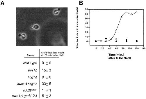
Figure 6.
(A) Mislocalization of nuclei in cells that are unable to delay in G2. Cells were treated as described in Figure 3. 4,6-Diamidino-2-phenylindole was used to stain the nuclei (swe1Δ hog1Δ cells shown). The same samples were also used to determine mitotic indices (Figure 3). The percentage of cells with separated nuclei that had not properly segregated (both nuclei in the mother cell) was determined ±SD from a minimum of three independent experiments. (B) Nuclear segregation is abnormal in hog1Δ cells following salt stress after release from alpha mating factor. Wild-type and hog1Δ cultures were arrested in G1 with alpha factor and released into fresh media. After 100 min, the culture was stressed by the addition of 0.4 M NaCl. The appearance of mislocalized nuclei in budded cells following the addition of NaCl was plotted versus time. A representative experiment with wild-type cells with or without salt (○ and ●, respectively) and hog1Δ cells with and without salt (▵ and ▴, respectively) is shown here.

