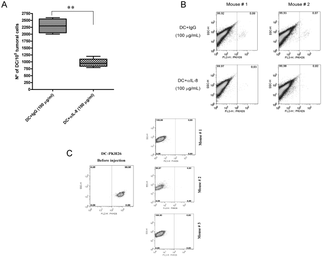
Figure 2. IL-8 produced by tumor cells in vivo retains DC inside xenografted tumor nodules.
(A) HT29 was xenografted into Rag−/− IL-2Rγ−/− double KO mice. Tumor nodules, 8–12 mm in diameter, were injected with 5×10⁶ PKH26-labeled monocyte-derived DC. When indicated, the 100 µL DC suspensions contained 100 µg/mL of mouse IgG (control antibody) or anti-IL-8 neutralizing mAb. The figure shows the proportion of PKH26+ events with respect to total tumor cells upon FACS analysis, three days after DC injection. Four mice with two bilateral xenografted tumors each per condition were used. Of note, DC cultured in the presence of the anti-IL-8 mAb at 100 µg/mL did not show loss of viability at least in 72 h (data not shown). (B) Representative FACS dot plots from A in two tumor nodules from two mice are shown as an example. Similar data were obtained with xenografts of the CaCo2 cell line (Figure S2). (C) Absence of PKH26-labeled DC in 3 out of 3 SW48 xenografts processed as in A, following injection of fluorescence labeled human DC. In the left dot-plot, the fluorescence intensity of injected PKH26-labeled DC is shown for reference. Dot plots are from representative experiment of two actually performed with three animals per group each.

