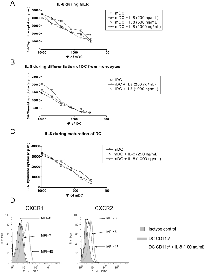
Figure 3. IL-8 does not impair T cell stimulation by DC that express functional CXCR1 and CXCR2.
(A) Human monocyte-derived DC and T-cells were seeded in the indicated proportions. Functional recombinant IL-8 was added at different concentrations as indicated in the graph legends. T-cell proliferation was measured by 3H-thymidine incorporation 3 days later. A representative case out of three independently performed experiments with cells from different combinations of donors is shown. (B) Experiments as in A, but in this case DC were incubated with the indicated amounts of IL-8 added to monocytes during the 7-day differentiation culture in the presence of GM-CSF+IL-4. A representative experiment out of at least three is shown. (C) Similar experiments as in B but in this case IL-8 at the indicated concentrations was added during the maturation 48 h culture onto differentiated DC matured for 48 h with IFN-α, TNF-α and poly I:C. A representative experiment out of at least three performed is shown. As a control every IL-8 batch was shown to readily attract human PMNs in chemotaxis assays (Figure S3). (D) Mature DC as those used for the MLRs were tested by indirect immunofluorescence for the expression of CXCR1 and CXCR2. Incubation with 100 ng/mL of IL-8 during 2 h resulted in a loss of fluorescence intensity upon immunostaining of the surface receptors indicating receptor internalization. The mean fluorescence intensity (MFI) of each histogram is provided. FACS Experiments were repeated in two occasions with similar results.

