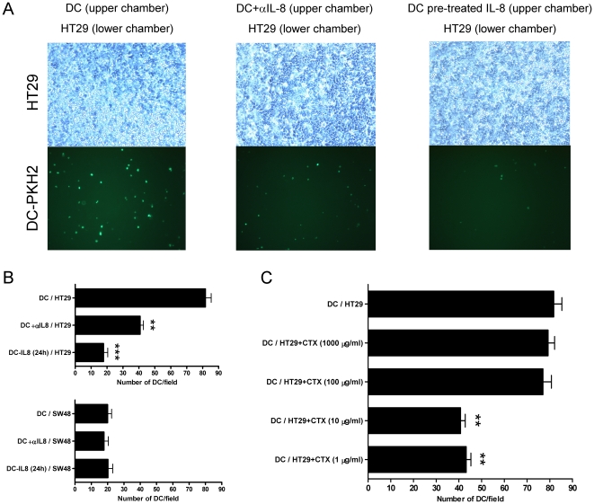
Figure 5. DC pre-exposed to IL-8 become desensitized to respond to carcinoma-derived IL-8 as a chemoattractant.
(A) Chemotaxis assays were set up with HT29 confluent monolayers in the lower chamber and fluorescent DC in the upper chamber. Phase contrast microscopy images and the corresponding UV fluorescence microscopic fields of the lower chamber are shown. When indicated the lower chamber contained neutralizing anti-IL-8 mAb (20 µg/mL) or the DC had been pre-exposed for 24 h to recombinant IL-8 (1 µg/mL). (B) In the upper panel representation of data from three independent experiments similarly performed to those in A with HT29 cells represented as mean±SD in which four random fields were counted for each triplicate well. In the lower panel experiments performed as in A, but in this case the confluent monolayers in the lower chamber were formed by SW48 cells that do not produce IL-8. (C) Experiments as in A, but in this case HT29 cells had been pretreated with various cyclophosphamide concentrations for 24 h as indicated in the figure. Results represent mean±SD.

