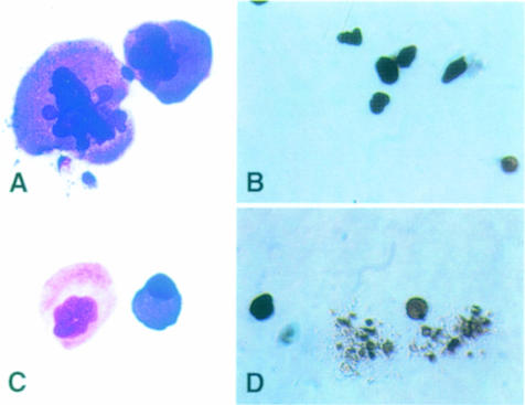
Fig. 3. Megakaryocyte morphology and in vitro platelet formation potential in mafG+/–::mafK+/– and mafG–/–::mafK+/– mice. Megakaryocytes were isolated from the bone marrow of mafG–/–::mafK+/– mice (A and B) or mafG+/–::mafK+/– mice (C and D). The morphology of both was examined by Giemsa staining (A and C). After plating into culture dishes, control megakaryocytes formed proplatelet projections after 24 h in culture (D), while megakaryocytes recovered from mafG–/–::mafK+/– mutants showed no propensity for megakaryocyte fragmentation or proplatelet formation (B).

An official website of the United States government
Here's how you know
Official websites use .gov
A
.gov website belongs to an official
government organization in the United States.
Secure .gov websites use HTTPS
A lock (
) or https:// means you've safely
connected to the .gov website. Share sensitive
information only on official, secure websites.
