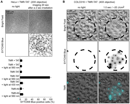
Figure 2. The photolytic and photocytotoxic effects of TMR-TAT occur rapidly and in all irradiated cells.
A) Irradiation of TMR-TAT endocytosed by cells causes plasma membrane blebbing and permeabilization (SYTOX® Blue staining) within 20 seconds after 2 sec irradiation at 560 nm(the radiant exposure is approximately 40 J/cm2). Cell destruction is observed in more than 95% of the cells irradiated at 560 nm. In contrast, no photocytoxicity is observed when cells treated with TMR-K9 (10 µM) or TMR (3 µM) and TAT (3 µM) are irradiated under similar conditions. Cells incubated with TMR-TAT but irradiated at 430 nm are not destroyed (excitation wavelength of SYTOX® Blue but not of TMR, radiant exposure was also approximately 40 J/cm2). The histogram represents the average percentage of cells stained by SYTOX® Blue after peptide and light treatment (the number of cells examined is at least 3000) and the error bars represent the standard deviation (experiments were reproduced at least 3 times). B) The cytotoxic effect of TMR-TAT is limited to only irradiated areas. COLO 316 cells were incubated with TMR-TAT (3 µM) for 1 h. The cells were washed with fresh media and endocytosis of TMR-TAT was confirmed by fluorescence microscopy. The cells within the circled area were exposed to light at 560 nm. Morphological changes and SYTOX® Blue staining (inverted monochrome or pseudo-colored cyan) are only observable in the irradiated area.

