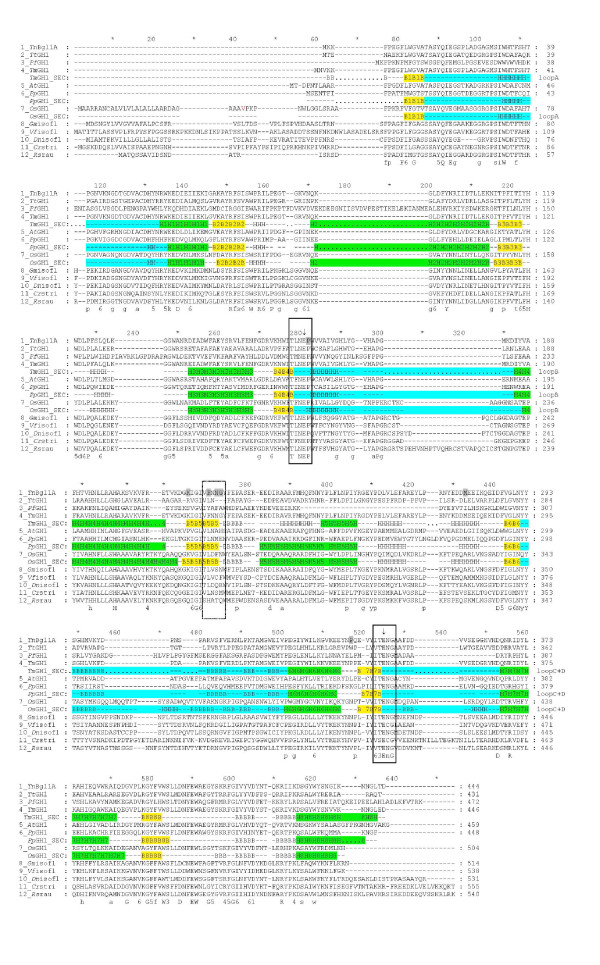
Figure 3.
Multiple sequence alignment of GH1 representatives. A multiple sequence alignment of β-glucosidases and flavonoid glucosidases from GH1. TnBgl1A, Thermotoga neapolitana Bgl1A (this work); TmGH1, Thermotoga maritima BglA (Q08638); AtGH1, Agrobacterium tumefaciens glucosidase (Q7CV27); PpGH1, Paenibacillus polymyxa BglB (P22505); OsGH1, Oryza sativae (rice) glucosidase (Q42975); GmGH1, Glycine max (soy) isoflavonoid glucosidase (AB259819); Vfiso, Viburnum_furcatum isoflavonoid glucosidase (AB122081); Dniso, Dalbergia nigrescens isoflavonoid glucosidase (AY766303); Crstri, Catharanthus roseus strictosidine β-glucosidase (Q9M7N7); Rsrau, Rauvolfia serpentina raucaffricine β-glucosidase (Q9SPP9). The region selected for mutagenesis is marked by a dashed box, and the two conserved motifs are boxed. The catalytic residues are indicated by arrows. Mutated residues are shaded in grey. Secondary structures are indicated below structure determined enzymes. Helices and strands of the β/α8-barrel are numbered and indicated in green and yellow, respectively. The sequence parts corresponding to the four loops (A-D) are indicated in cyan. A consensus sequence is shown in bold below the aligned sequences. Completely conserved resides are shown in upper cases, residues conserved in more than 80% of the sequences are shown in lower case, positions with related residues are indicated by numbers (1 = N, D; 3 = S, T; 4 = K, R; 5 = Y, F, W; 6 = I, L, V, M).

