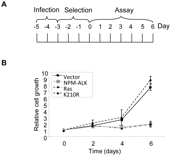
Figure 1. NPM-ALK expression in primary MEFs inhibits cellular proliferation.
(A) Experimental design and reference timeframe. Infection refers to exposure of MEFs to retrovirus, and selection refers to enrichment for transduced cells with 2 µg/ml of puromycin. (B) MEFs infected with retrovirus and selected for expression of the indicated genes were assessed for their ability to proliferate over 6 days via crystal violet assay. Relative cell growth was determined by comparing crystal violet optical density values obtained at 590 nm at each time point with those obtained at day 0. Each time point was conducted in triplicate in at least 3 separate experiments using MEFs from independent embryo preparations. Error bars are standard deviations of the mean.

