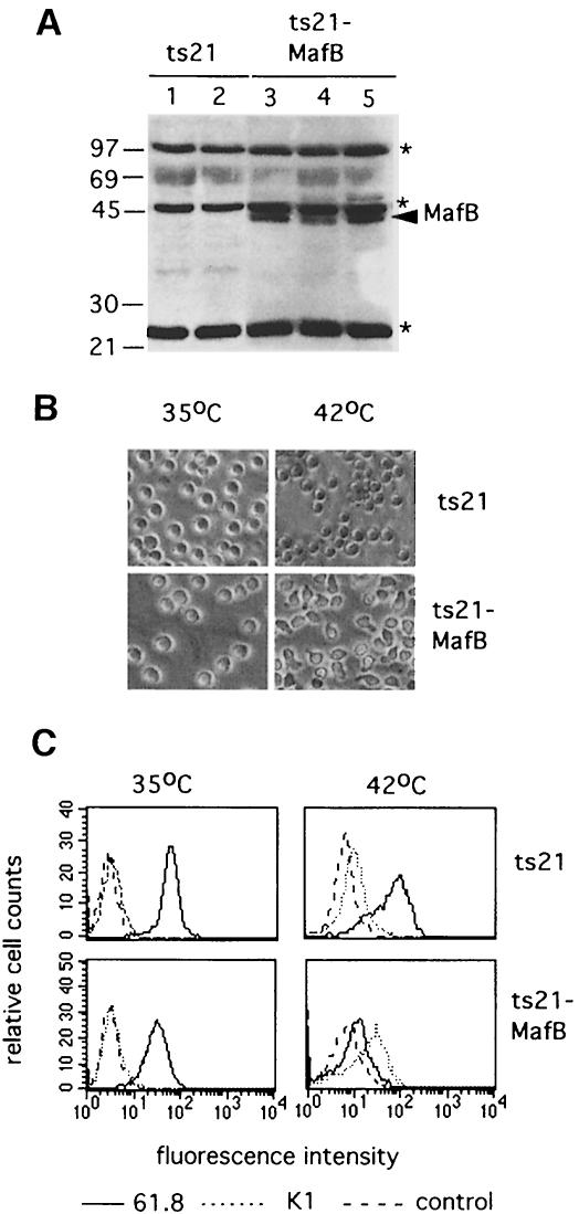
Fig. 5. (A) Western blot of cellular extracts from myeloblast clones expanded from ts21- and ts21-MafB-transformed colonies using a monoclonal antibody directed against the HA epitope tag in the ts21-MafB construct. Two ts21 control clones and three ts21-MafB clones are shown. The arrowhead indicates MafB protein, the asterisks indicate background bands detected in all samples. The size of molecular weight standards is shown on the left. (B) Phase contrast photomicrograph of a representative ts21 and ts21-MafB clone each maintained at 35°C or shifted to 42°C for 48 h. (C) FACS profiles of representative ts21 and ts21-MafB clones maintained at 35°C or shifted to 42°C for 48 h using the myeloblast-specific antibody 61.8 (–––) the macrophage antigen-detecting antibody K1 (- - - -) and an isotype-matched control antibody against an intracellular antigen (— — —).

An official website of the United States government
Here's how you know
Official websites use .gov
A
.gov website belongs to an official
government organization in the United States.
Secure .gov websites use HTTPS
A lock (
) or https:// means you've safely
connected to the .gov website. Share sensitive
information only on official, secure websites.
