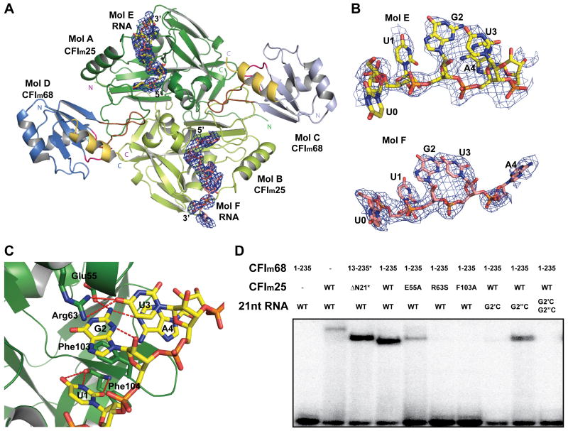
Figure 3.
The CFIm complex binds two UGUA elements simultaneously.
(A) Overview of the CFIm-UAUUUUGUA complex. RNA molecules are shown as stick model and colored in yellow and salmon. Simulated annealing omit map for the RNA molecules is shown as a blue mesh and contoured a 2.5σ. (B) A close up view of the bound RNA molecules. Density was observed for the UGUA elements and the ribose of the base preceding the first U (U0). (C) A close up view of CFIm25 interacting with UGUA elements in Mol A. Residues participating in RNA binding are shown in stick model representation. Hydrogen bonds are represented by red dashed lines. (D) Electrophoretic mobility shift assays (EMSA) of CFIm variants in complex with various PAPOLA RNA sequence variants. An asterisk (*) indicates that the protein underwent reductive methylation. A single prime (′) represents the first UGUA element, and double prime (″) represents the second UGUA element. See also Figure S3.

