
Fig. 2. Confirmation of differential expression of SA-miRNAs in senescent cells.
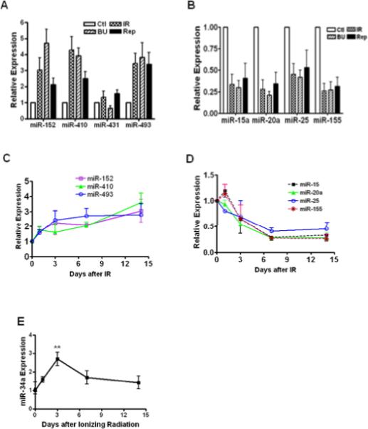
TaqMan MicroRNA Assays were used to determine the expression levels of SA-miRNAs in senescent WI-38 cells. The levels of up-regulated (A) and down-regulated (B) SA-miRNAs in replicatively senescent cells (Rep) and prematurely senescent cells (induced by IR and BU) were normalized to the control cells and graphed. Kinetic changes of up-regulated (C) and down-regulated (D) SA-miRNAs versus time after IR were plotted. Kinetic changes of miR-34a expression were determined (E). Data are presented as fold changes compared with control (Mean ± SE, n = 4). **, p < 0.01 vs. control.