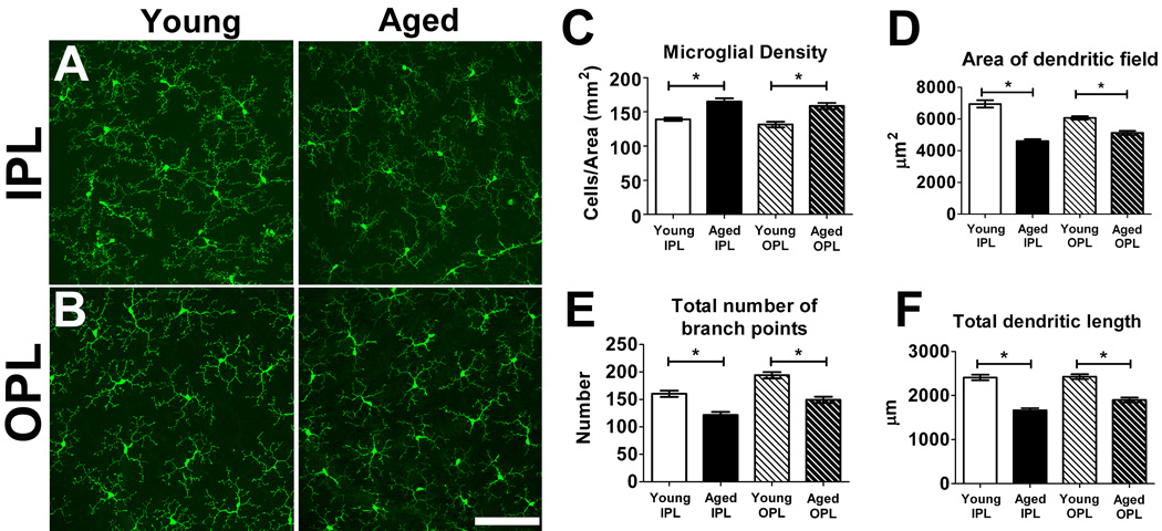
Figure 1. Morphology and distribution of retinal microglia in young (2–3 months old) and aged (18–24 months old) CX3CR1+/GFP mice.
Resting retinal microglia, as visualized in retinal flat-mount preparations, possess small somata and ramified morphologies that were regularly distributed in the vitreal half of the retina with processes located in the inner plexiform layer (IPL) (A) and the outer plexiform layer (OPL) (B). Qualitatively similar microglial distributions and morphologies were observed in both young (left panels) and aged (right panels) retinas. Scale bar = 100 µm. (C) Densities of microglial somata in both IPL and OPL layers of young and aged retinas were measured in the mid-peripheral retina, showing increased microglial cell densities in the IPL and the OPL in aged compared to young retina (n = 24 retina from 6 young and 6 aged animals). Analyses of morphological parameters of area of dendritic field (D), total number of branch points (E), and total dendritic length of individual microglia (F), revealed that aged microglia have a smaller and less branched dendritic morphologies compared to young microglia in both the IPL and the OPL layers (n = 255 cells from 6 young animals and 205 cells from 6 aged animals). (Asterisks indicate comparisons for which p<0.05)

