Figure 6.
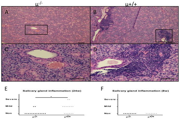
Salivary gland inflammation. A–D. Representative histology showed inflammatory cell infiltrates around the salivary gland ducts in B cell competent mice whereas no inflammation was observed in B cell deficient mice. E. Degree of salivary gland inflammation was significantly lower in B cell deficient mice at 24 weeks of age (μ−/−, n=15; μ+/+, n=15). F. Salivary gland inflammation was not observed in either group at 8 weeks of age (μ−/−, n=8; μ+/+, n=7). (H&E staining. Scale bar 100 μm. *: p < 0.05 in Mann-Whitney Test)

