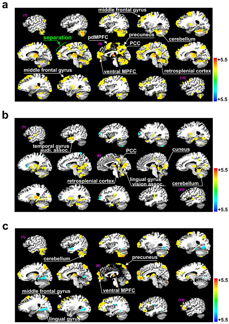
Fig. 2.

Brain regions demonstrating significant specific thalamic functional connections. Regions of particular interest are highlighted by white arrows. (a) Brain regions identified by one-sample t-tests with significant thalamic functional connectivity in healthy controls (Z-sore, p<0.025 after correction for multiple comparisons for here and elsewhere). (b) The same for VS patients. (c) Brain regions identified by two-sample t-tests (p<0.025) for a significant difference in specific thalamic functional connectivity between healthy subjects and VS patients.
