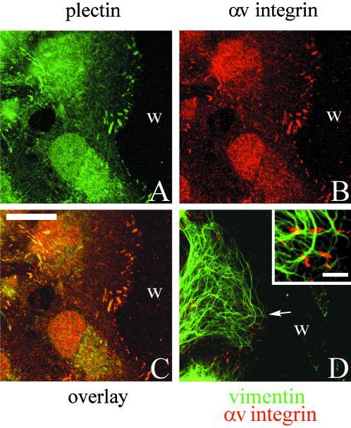
Figure 13.
A confluent monolayer of TrHBMECs was wounded by scraping with a pipette-man tip. The culture was allowed to recover for 4 h, at which time cells had begun to migrate into the wound area. The preparation was then processed for double-label immunofluorescence microscopy using antibodies against plectin (A) together with an antiserum against αv-integrin (B). Cells were viewed by confocal microscopy. The focal plane is close to the substratum-attached surface of the cells. C is the overlay of the two fluorescence images. Preparations were also made of wounded cultures for double-label immunofluorescence using the antiserum against the αv-integrin (red) combined with a monoclonal vimentin antibody (green). The overlay of the two images is shown in D. Inset in D shows a higher power view of several αv-integrin positively stained focal contacts with which vimentin interacts in a cell that has migrated into the wound site (w). Bar in C, 20 μm; bar in D, inset, 3 μm.

