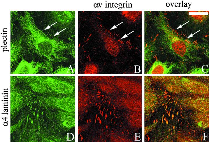
Figure 3.
TrHBMECs were maintained on glass coverslips until the cells reached confluence. Approximately 24 h later the cells were processed for double-label immunofluorescence microscopy using an antibody against plectin (A) or 2A3 antibody against the α4 laminin subunit (D) in combination with an antiserum against αv-integrin (B and E). Cells were viewed by confocal microscopy. The focal plane is close to the substratum-attached surface of the cells. C and F show the overlays of the fluorescence images. Arrows in A–C indicate areas of focal contacts stained by αv-antibodies in B that are not recognized by plectin antibodies in A. Rather, the plectin antibodies in A generate a predominantly filamentous staining pattern. Bar in C, 20 μm.

