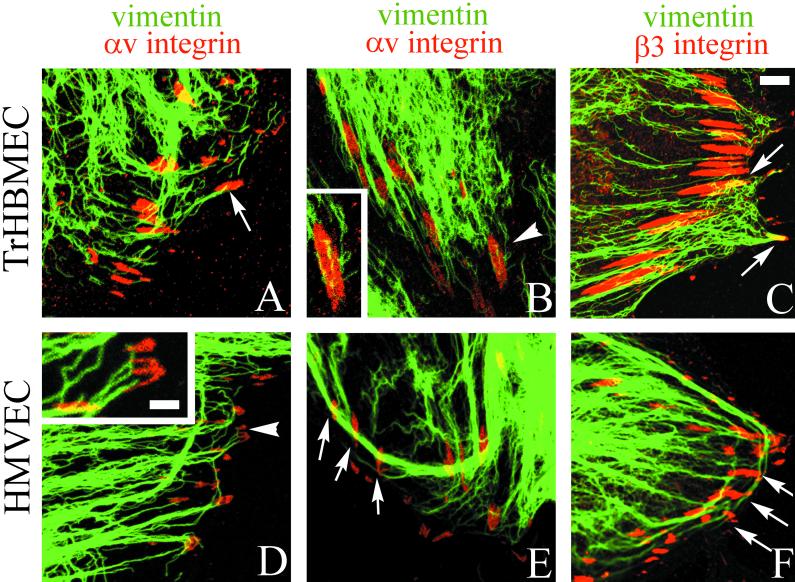
Figure 6.
TrHBMECs (A–C) and HMVECs (D–F) were processed for double-label immunofluorescence using a monoclonal antibody against vimentin (green) in combination either with an antiserum against the αv integrin subunit (red) (A, B, D, and E) or an antiserum against β3 integrin (red) (C and F). Overlays of the staining patterns are shown. In TrHBMECs some vimentin filament bundles can be seen to terminate at αv-integrin–containing focal contacts (arrow in A). The focal contact indicated by the arrowhead in B is shown at higher magnification in the inset. Vimentin filaments appear to be wrapped around the focal contact. In HMVECs vimentin bundles are also seen in association with αv-integrin–containing focal contacts (arrowhead and arrows in D and E). The inset in D shows a higher power view of one focal contact (arrowhead) in the HMVEC cell shown in D. Note that three individual vimentin filament bundles appear to terminate at the site of the focal contact. Vimentin filaments and bundles also associate with focal contacts stained by β3 integrin antibodies in both TrHBMECs and HMVECs (arrows in C and F). Bar in C, 5 μm; bar in D (insets), 1 μm.

