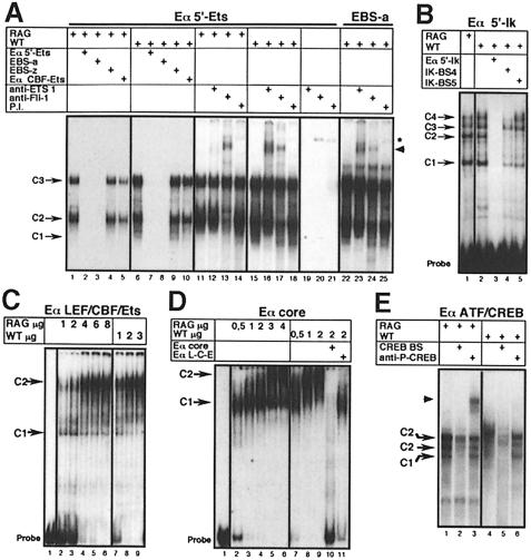
Fig. 6. EMSA analysis of Eα factor binding sites. Radiolabeled probes corresponding to different Eα regions (indicated at the top of each panel) were used together with RAG–/– (RAG) or wild-type thymocyte nuclear extracts (NE), as indicated. Unlabelled oligonucleotides used for competition analyses (A, B, D and E), as well as anti-sera used for supershift analyses (A and E), are also indicated. P.I., pre-immune rabbit serum. The position of the major complexes observed with the individual probe are indicated by arrows (C#; see details in the text). Supershifts are indicated by arrowheads. In (A), lanes 1–10 and 11–25 correspond to two separate experiments; a longer exposure is presented for lanes 11–14 to visualize the shifted bands better. The star indicates a non-specific complex that can be formed in the absence of NE. Probes and oligonucleotide competitors are defined in Table I.

An official website of the United States government
Here's how you know
Official websites use .gov
A
.gov website belongs to an official
government organization in the United States.
Secure .gov websites use HTTPS
A lock (
) or https:// means you've safely
connected to the .gov website. Share sensitive
information only on official, secure websites.
