
Fig. 6.
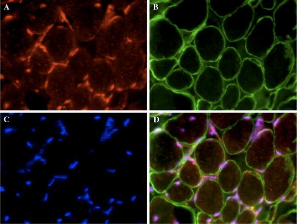
ZBTB42 localizes to the nuclei of human skeletal muscle fibers and merges with a nuclear stain. Immunofluorescence analysis of a triple-stained muscle section shows nuclear localization of ZBTB42 in skeletal muscle fibers. Sections are cut laterally through the muscle resulting in cross-sectional view of the muscle cells. Muscle cell nuclei are peripherally localized. a AX-1#3 stain of ZBTB42 in human skeletal muscle shows staining exclusive to the nuclei (Cy3-conjugated secondary antibody). b Anti-merosin staining shows expression of the membrane protein, merosin (Cy2-conjugated secondary antibody). c DAPI nuclear stain. d Merge of all three stains shows perfect alignment of DAPI and ZBTB42 expression in muscle cell nuclei. Images were taken on an apotome microscope at 40× magnification