
Figure 3.
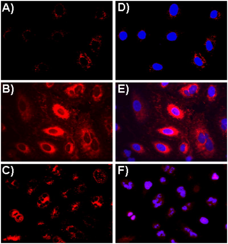
Assessment of IONPs’ cellular uptake via confocal laser-scanning microscopy using lung carcinoma A549 cells. A) No internalization was observed in cells treated with carboxylated IONPs (2), as no DiI fluorescence was observed in the cytoplasm, B) Enhanced internalization was observed upon incubation with the folate-immobilized IONPs (4), C) Cells incubated with Taxol and DiI co-encapsulating folate-functionalized IONPs (5) induced cell death. (D–F) Corresponding merged confocal images of the functional IONPs treated cells with their nucleus stained with DAPI (blue).