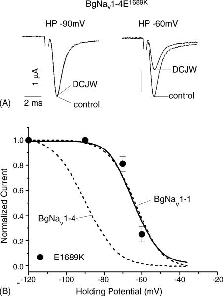
Fig. 6.
Inhibition of peak current of BgNav1-4E1689K by DCJW. (A) Sodium currents recorded by 20 ms depolarization to −10 mV from the holding potential of −90 or −60 mV in control or in the presence of DCJW (20 μM) for 30 min. (B) Voltage-dependence of DCJW inhibition. The protocols were the same as those in Fig. 2. Voltage-dependence of DCJW inhibition of BgNav1-1 and BgNav1-4 from Fig. 2 is indicated in dash lines for direct comparison. Each point represents data from four oocytes.

