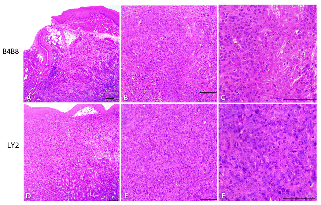
Figure 4.
Representative hematoxylin and eosin stained light microscopy images showing the phenotypic features of B4B8 and LY2 primary tumors. Low power (40×) views (A & D) of these tumors reveal invasive islands and cords of malignant squamous cell carcinomas underneath the host’s oral mucosal epithelium (bar = 200 µM). At higher magnifications (100X & 200×), B4B8 tumor (B &C) and LY2 tumors (E & F) reveal histopathologic features of well-differentiated low-grade and poorly-differentiated high-grade squamous cell carcinomas, respectively (bar = 100 µM).

