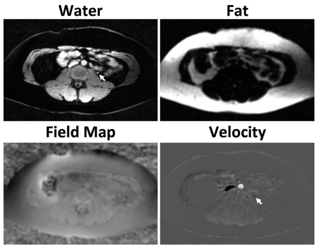
FIG. 9.
Example axial images obtained in abdominal scans of a healthy volunteer, using the proposed CSI-PC procedure. Velocity images are masked by the water magnitude image for improved visualization. The results demonstrate excellent separation of fat and water. Field map images from CSI-PC show a minimal influence from fat, likely caused by inaccurate characterization of the multiple peaks of fat. This allows for B0 heterogeneity corrections without losing small vessels embedded in fat (arrows).

